42 onion cells under microscope with labels
Onion Epidermal Cell Labeled Diagram - schematron.org Draw a labelled diagram of an onion epidermal cell seen under the microscope. ( 4 marks) e The onion epidermal cells are not green in colour because they lack. The epidermal cells of onions provide a protective layer against viruses and fungi that may harm the sensitive tissues. rhoeo discolor leaf under microscope labeled - Erotske priče In contrast, the light has to pass through the specimen to form the image under a compound microscope. Cutter 6. No need to register, buy now! Zebrina. Take an onion bulb/ rhoeo leaf, with the help of forceps pull a thin transparent peel. In Microscope Lab II, we look at the __ of a leaf of the Rhoeo discolor plant to see representative plant cells.
Onion cells under the microscope: 40X - 100X - 400X - YouTube under the #microscope: 40X - 100X - 400X
Onion cells under microscope with labels
Required practical - using a light microscope - Cells in ... - BBC Bitesize Record the microscope images using labelled diagrams or produce digital images. When first examining cells or tissues with low power, draw an image at this stage, even if going on to examine the ... Onion Cells Under a Microscope - Requirements/Preparation/Observation Add a drop of iodine solution on the onion membrane (or methylene blue) Gently lay a microscopic cover slip on the membrane and press it down gently using a needle to remove air bubbles. Touch a blotting paper on one side of the slide to drain excess iodine/water solution, Place the slide on the microscope stage under low power to observe. Microscopy, size and magnification - Microscopy, size and ... - BBC Place cells on a microscope slide. Add a drop of water or iodine (a chemical stain). Lower a coverslip onto the onion cells using forceps or a mounted needle. This needs to be done gently to...
Onion cells under microscope with labels. Observing Onion Cells Under The Microscope Afterwards, carefully mount the prepared and stained onion cell slide onto the microscope stage. Make sure that the cover slip is perfectly aligned with the microscope slide, and that any excess stain has been wiped off. Secure the slide on the stage using the stage clips. Labeled Onion Cell Under Microscope 40x - Micropedia Labeled onion cell under microscope 40x. While photosynthesis takes place in the leaves of an onion containing chloroplast the little glucose that is produced from this process is converted in to starch starch granules and stored in the bulb. This slide was scanned using a 40x 080na objective. Machine vt recommended for you. PDF Onion Cell Lab - SomeWaresInMaine Research Biology Onion Cell Lab page 1 of 3 Onion Cell Lab After you have completed the rest of this lab come back to this cover page DRAW & LABEL AN ONION CELL WITH ALL THE PARTS / ORGANELLES YOU OBSERVE UNDER 40X. Purpose: To observe and identify major plant cell structures and to relate the structure of the cell to its function. animal cell under microscope labeled - Rayford Runyon When observing onion cells there is the Cell Surface Membrane which is present in all living cells. Most cells both animal and plant range in size between 1 and 100 micrometers and are thus visible only with the aid of a microscope. While observing with tissues or on tissue.
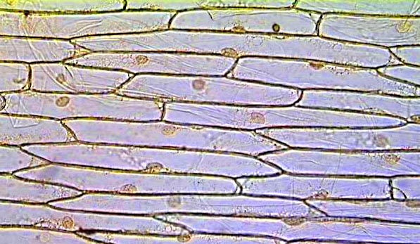
Animal Cell Under Light Microscope Labelled : Draw and label the ... Onion cell diagram labeled structure of animal cell and plant cell under microscope. An organelle found in large numbers in most cells, in which the biochemical processes of respiration and energy production occur. Under a light microscope, the cell membrane, nucleus and cytoplasm of a cheek cell (animal cell) can be observed. Observing Onion Cells under a Microscope - Blog, She Wrote you'll need to stain the onion cells before you observe them under the microscope. There are different types of stains depending on what type of cell you are going to look at. Iodine - dark stain that colors starches in cells. In an onion cell, it will make the cell wall more visible. It provides some contrast for viewing under a microscope. PDF Onion Cells - Investigation - Exploring Nature 1. Observe the onion tissue under the microscope at 4x, 10x and 40x with lots of light (open diaphragm). 2. Then slowly close the diaphragm while observing the image to find the best light for seeing cellular details. 3. Draw a section of onion skin cells at 10x magnification. 4. Switch to 40x and draw one cell and label it. Microscope Cell Lab: Cheek, Onion, Zebrina - SchoolWorkHelper The onion epidermis cell is the only cell that has a cell wall. In addition, it is the only cell that has a chloroplast, where photosynthesis can happen. The cheek epithelium cell is the only one that has centrioles, the barrel-shaped organelle that is responsible for helping organize chromosomes during cell division.
Blog, She Wrote - Embracing the Independent & Authentic Nature of ... Blog, She Wrote - Embracing the Independent & Authentic Nature of ... Onion Cells Microscope Stock Photos and Images - Alamy Onion cells under the microscope. Garden onion, Bulb Onion, Common Onion (Allium cepa), cell tissue of a garden onion with dyed chromosomes, light microscopy, x 120, Germany. Onion Cells under the Microscope. Onion skin cells under the microscope, horizontal field of view is about 0.61 mm. Detailed view of the cells of a red onion as seen ... Animal Cell Diagram Under Microscope Labeled Animal Cell Diagram Under Microscope Labeled Sunday, April 18th 2021. | Diagram Animal Cell Diagram Under Microscope. Function cell does in the body dictate the change and adaptation done by cell. When observing onion cells, there is the Cell Surface Membrane which is present in all living cells. Onion Cell Lab Report.docx - Onion Cell Lab Report By station, remove the single layer of epidermal cells from inner side of the scale leaf. 3(Place the single layer of onion on a glass slide. 4(Place a drop of iodine stain on your onion tissue. 5(Put the cover slip on the stained tissue and gently tap out any air bubbles. 6(Observe the cells under the microscope and see you results.
PPT - The Light Microscope PowerPoint Presentation, free download - ID:2332804
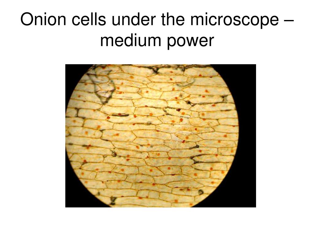
DOC The Onion Cell Lab - chsd.us Onion tissue provides excellent cells to study under the microscope. The main cell structures are easy to see when viewed with the microscope at medium power. For example, you will observe a large circular . nucleus. in each cell, which contains the genetic material for the cell. In each nucleus, are round bodies called . nucleoli

Microscope Onion Cell Labeled - Micropedia
Onion Root Tip Mitosis - Stages, Experiment and Results · Cover the sample (root tip) with a coverslip and gently press the coverslip down, then examine the slide under the microscope starting with low magnification * For this experiment, a properly prepared slide should appear light pink due to the stain to almost colorless. * Unused roots can be stored in 70 percent alcohol. Results

Rens blog : Science, cells
Onion Skin Cells - Investigation - Exploring Nature 5. Observe the onion tissue under the microscope at 4x, 10x and 40x with lots of light (open diaphragm). Then slowly close the diaphragm while observing the image to find the best light for seeing cellular details. 6. Draw a section of onion skin cells at 10x magnification. Then switch to 40x and draw one cell and label it.

onion cells under microscope - YouTube
Onion Skin Epidermis Sample under microscope 4x,10x Magnification A sample on an onion skin epidermis diyed in blue for visibility, viewd under the microscope at 4x and 10x magnification.microscope:Biolux model :AL
Red Onion Cell Under Microscope Labeled - Micropedia
Onion Root Mitosis - Microscopy-UK It is common to see photomicrographs of onion root cells when demonstrating how cell division takes place in plants. Onions have larger chromosomes than most plants and stain dark. The chromosomes are easily observed through a compound light microscope. The cells pictured below are located in the apical meristem of the onion root. The apical ...
Cell Biology Gifts on Zazzle AU
Looking at the Structure of Cells in the Microscope Both types of light microscopy are widely used to visualize living cells. Figure 9-7 Two ways to obtain contrast in light microscopy. (A) The stained portions of the cell reduce the amplitude of light waves of particular wavelengths passing through them. A colored image of the cell is thereby obtained that is visible in the ordinary way. (more...)
Why can't we see cells? - Science with Mrs Pizzimenti
DOC Plant and Animal Cells Microscope Lab - hillsboro.k12.oh.us Make a drawing of one onion cell, labeling all of its parts as you observe them. (At minimum you should observe the nucleus, cell wall, and cytoplasm.) Cheek cells 1. To view cheek cells, gently scrape the inside lining of your cheek with a toothpick. DO NOT GOUGE THE INSIDE OF YOUR CHEEK! (We will observe blood cells in a future lab!!) 2.

Rens blog : Science, cells
Biology Experiment Examination of Onion Cell in Light Microscope Place the single layer of onion cell epithelium on a glass slide. Make sure that you do not fold it over or wrinkle it. Place a drop of iodine stain on your onion tissue. Put the cover slip on the stained tissue and gently tap out any air bubbles. Observe the cells under 4x, 10x, and 40x with the diaphragm wide open.

Onion Epidermal Cell Labeled - Top Label Maker
Onion Epidermis - kuensting.org Onion epidermis at 40X, iodine stain. Onion epidermis, at 100X, iodine stain. Onion epidermal cells, iodine stain, 400X. The nucleus of an onion epidermal cell, 1000X magnification.

Onion Cells Under Microscope | Biology - YouTube
Microscopy, size and magnification - Microscopy, size and ... - BBC Place cells on a microscope slide. Add a drop of water or iodine (a chemical stain). Lower a coverslip onto the onion cells using forceps or a mounted needle. This needs to be done gently to...

Life Science Unit - WELCOME TO MR.FLEMING SCIENCE
Onion Cells Under a Microscope - Requirements/Preparation/Observation Add a drop of iodine solution on the onion membrane (or methylene blue) Gently lay a microscopic cover slip on the membrane and press it down gently using a needle to remove air bubbles. Touch a blotting paper on one side of the slide to drain excess iodine/water solution, Place the slide on the microscope stage under low power to observe.

Labeled Onion Cell Under Microscope 40x - Micropedia
Required practical - using a light microscope - Cells in ... - BBC Bitesize Record the microscope images using labelled diagrams or produce digital images. When first examining cells or tissues with low power, draw an image at this stage, even if going on to examine the ...

Rens blog : Science, cells

Lab activities - YongQian's e-portfolio
How many onion skins are there?

Light Microscope Onion Cell Labeled - Micropedia

BIOLOGYBUDDY13
Post a Comment for "42 onion cells under microscope with labels"