43 diagram of the brain with labels and functions
Brain (Human Anatomy): Picture, Function, Parts, Conditions, and More • The cerebellum is at the base and the back of the brain. The cerebellum is responsible for coordination and balance. The brain is also divided into several lobes: • The frontal lobes are... Human Brain: Structure, Location, Function, Parts & Pictures Spinal cord is the central nervous system component that begins in the lower area of the brain, extending along the spine. The spinal cord links the brain with the nerves. The spinal cord's nerve tissues are approximately 45 centimeters long, and nearly 2 centimeters bulk, and they conform the peripheral nervous system. Function of the Brain
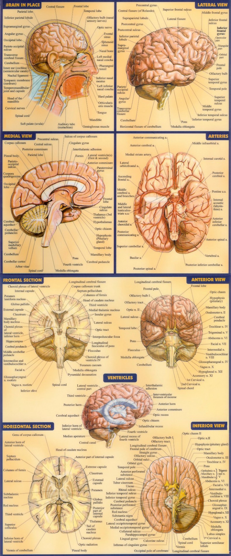
Parts of the brain: Learn with diagrams and quizzes | Kenhub Labeled brain diagram First up, have a look at the labeled brain structures on the image below. Try to memorize the name and location of each structure, then proceed to test yourself with the blank brain diagram provided below. Labeled diagram showing the main parts of the brain Blank brain diagram (free download!)

Diagram of the brain with labels and functions
developingchild.harvard.edu › resources › the-brainThe Brain Circuits Underlying Motivation: An Interactive Graphic The brain systems that govern motivation are built over time, starting in the earliest years of development. These intricate neural circuits and structures are shaped by interactions between the experiences we have and the genes we are born with, which together influence both how our motivation systems develop and how they function later in life. Structure, Diagram, Parts Of Human Brain - BYJUS The hypothalamus is a small and essential part of the brain, located precisely below the thalamus. It is considered the primary region of the brain, as it is involved in the following functions: Receives impulses Regulates body temperature Controls the mood and emotions Controls the sense of taste and smell Synthesises the body's essential hormones › photos › muscular-systemMuscular System Labeled Diagram Stock Photos, Pictures ... Cranial nerves vector illustration. Labeled diagram with brain sections. Cranial nerves vector illustration. Labeled diagram with brain sections and its functions with senses. Regions with olfactory, optic, abducent, facial and vagus parts. muscular system labeled diagram stock illustrations
Diagram of the brain with labels and functions. Ventricles of the Brain: Labeled Anatomy, Function, CSF Flow ... We will use labeled diagrams and lateral views of the brain to learn the anatomy, boundaries, and locations of each ventricle. We will also learn how cerebrospinal fluid is produced, and how it flows throughout the ventricular system, subarachnoid space, and central canal of the spinal cord. sfponline.org › Uploads › 71NERVOUS SYSTEM WORKSHEET - St. Francis Preparatory School 1. The diagram below is of a nerve cell or neuron. Add the following labels to the diagram: Axon Myelin sheath Cell body Dendrites Muscle fibers Axon terminals 2. Color in the diagram as suggested below. Axon - purple Axon Terminals – orange Myelin sheath – yellow Cell body – blue Dendrites – green Muscle fibers - red 3. byjus.com › biology › diagram-of-neuronA Labelled Diagram Of Neuron with Detailed Explanations A neuron is a specialized cell, primarily involved in transmitting information through electrical and chemical signals. They are found in the brain, spinal cord and the peripheral nerves. A neuron is also known as the nerve cell. The structure of a neuron varies with their shape and size and it mainly depends upon their functions and their ... Label The Brain Psychology Teaching Resources | Teachers Pay Teachers This bundle pack includes a 2-hour comprehensive presentation on the parts and functions of the brain. It also includes a student-guided notes document where they label a diagram of the brain. It also includes a master copy of that diagram for grading. Subjects: Psychology, Social Studies - History. Grades:
Anatomy of the Brain - Simply Psychology The temporal lobes are located on both sides of the brain, near the temples of the head, hence the name temporal lobes (Figure 5). The main functions of these lobes include understanding, language, memory acquisition, face recognition, object recognition, perception, and processing auditory information. PDF Brain Anatomy - Wou Below are listed the major anatomical regions / landmarks of the brain stem with their corresponding functions (Figure 7): REGION / LANDMARKFUNCTION Midbrain Region of brain stem between the diencephalon and pons; contains multiple fiber tracts running between higher and lower neural centers. Cerebral peduncle Parts of the Brain Activity for Kids, Brain Diagram, and Worksheets for ... Their are 2 brain function worksheets where your student will learn about the different parts of the brain your child will learn about are: FRONTAL LOBES - The frontal lobes control voluntary movement such as reasoning, planning, parts of speech and movement, emotions, and problem-solving It is fully developed by age 10. Human Brain Lesson for Kids: Function & Diagram - Study.com The Cerebrum. The biggest part of your brain is called the cerebrum. One thing your cerebrum does is control your muscles. It has two halves, one on the left and one on the right. The weird thing ...
Brain: Function and Anatomy, Conditions, and Health Tips It helps regulate many important functions, including motor and sensory functions, breathing, sneezing, and swallowing. Brain conditions There are hundreds of conditions that can affect the brain. Nervous System - Label the Brain - TheInspiredInstructor.com This brain part controls balance, movement, and coordination. (11) This brain part controls involuntary actions such as breathing, heartbeats, and digestion. (12) This part of the nervous system moves messages between the brain and the body. (13) This part of the cerebrum interprets and sorts information from the senses. (14) PDF Psychology Brain Structure/Anatomy and Function Psychology - Brain Structure/Anatomy and Function BRAIN FACTS Composition of the brain: 78% water, 12% lipids, 8% protein, 1% carbs, 2% soluble organics, and 1% salt ... Some products are also labeled incorrectly." Nicotine in e-cigarettes raise blood pressure. Compared to nonusers, users of e-cigarettes have a 71% higher risk of stroke, 59 ... DOC Brain Anatomy Function Cheat Sheet Memory (remembering and learning) Amygdala Emotion (aggression) rage, fear Kluecer& Bucy Lesion monkey brain Hypothalamus Regulates thirst, hunger, body temperature, sexual behavior (hormone release). Controls/regulates maintenance reflexes (eating), Homeostasis linked to emotion. Helps govern endocrines. Monitors glands. Controls hunger.
Lacrosse Stadium as an Animal Cell
byjus.com › biology › diagram-of-heartHeart Diagram with Labels and Detailed Explanation - BYJUS The diagram of heart is beneficial for Class 10 and 12 and is frequently asked in the examinations. A detailed explanation of the heart along with a well-labelled diagram is given for reference. Well-Labelled Diagram of Heart. The heart is made up of four chambers: The upper two chambers of the heart are called auricles.

Anatomy and Functions of the Brain Medical Illustration
en.wikipedia.org › wiki › DopamineDopamine - Wikipedia Dopamine is also synthesized in plants and most animals. In the brain, dopamine functions as a neurotransmitter—a chemical released by neurons (nerve cells) to send signals to other nerve cells. Neurotransmitters are synthesized in specific regions of the brain, but affect many regions systemically.

yhst-71986762869995_2239_55942590 (400×518) | Brain science, Brain anatomy, Brain facts
14 Informative Facts, Diagram & Parts Of Human Brain For Kids The brain weighs just about two to three pounds and appears like a walnut. The brain is comprised of three main regions — cerebrum, cerebellum, and brainstem (3). Let us discuss these parts and their functions in more detail (1) (3) (4). Cerebrum: The cerebrum is the largest part of the brain.
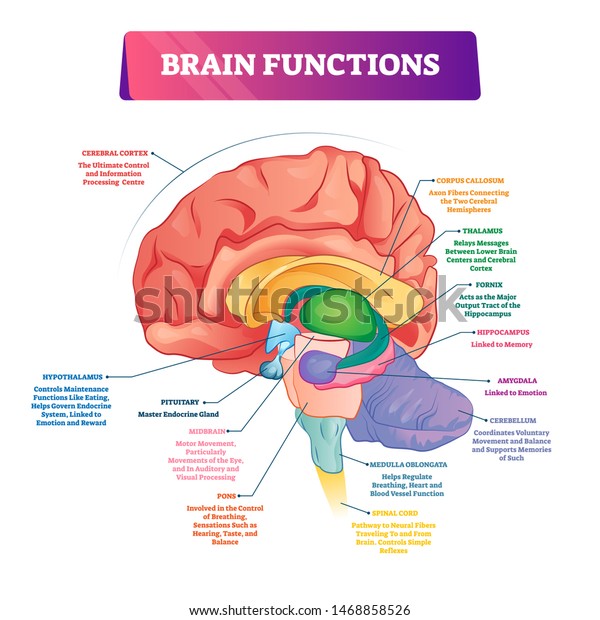
Brain Functions Vector Illustration Labeled Explanation Stock Vector (Royalty Free) 1468858526
external structure human heart diagram Drawing of the brain with labels. Labels brain drawing ear anatomy functions human diagram labeled clipartmag. Sheep dissection anatomy heart side dissected pig human class physiology card labeled valve science cow septum blood term cardiac interventricular ... labels brain drawing ear anatomy functions human diagram labeled clipartmag. Right ...

brain diagram 2 - /medical/anatomy/brain/brain_diagram_2.png.html
PDF The Human Brain Diagram - Therapist Aid Parietal Lobe • Interprets sensations, such as touch, pressure, pain, heat, and cold. • Helps with the understanding of objects, shapes, and space. Frontal Lobe • Suppresses socially inappropriate behavior. • Predicts consequences of actions. • Plays a role in the choice between helpful and harmful actions. Temporal Lobe

Educative diagrams: Digestive System Diagram
rsscience.com › stereo-microscopeParts of Stereo Microscope (Dissecting microscope) - Rs' Science If you would like to learn optical components of a compound microscope, please visit Compound Microscope Parts – Labeled Diagram and their Functions, and this article. How to use a stereo (dissecting) microscope. Follow these steps to put your stereo microscopes in work: 1. Set your microscope on a tabletop or other flat sturdy surface where ...

Pinterest • The world’s catalog of ideas
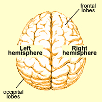
Lobes of the brain: Structure and function | Kenhub The lobes of the cerebrum are actually divisions of the cerebral cortex based on the locations of the major gyri and sulci. The cerebral cortex is divided into six lobes: the frontal, temporal, parietal, occipital , insular and limbic lobes. Each lobe of the cerebrum exhibits characteristic surface features that each have their own functions.

The Brains Functions by Crystal Montano and Mellanie Quijano - ThingLink
Diagram of the Brain and its Functions - Bodytomy Given below is a diagram outlining the main brain functions and parts. Vertical Section of the Brain and its Functions Midbrain The midbrain is divided into two parts by the Aqueduct of Sylvius, which is the duct that connects the IIIrd ventricle in the midbrain with the IV ventricle in the pons and medulla oblongata.
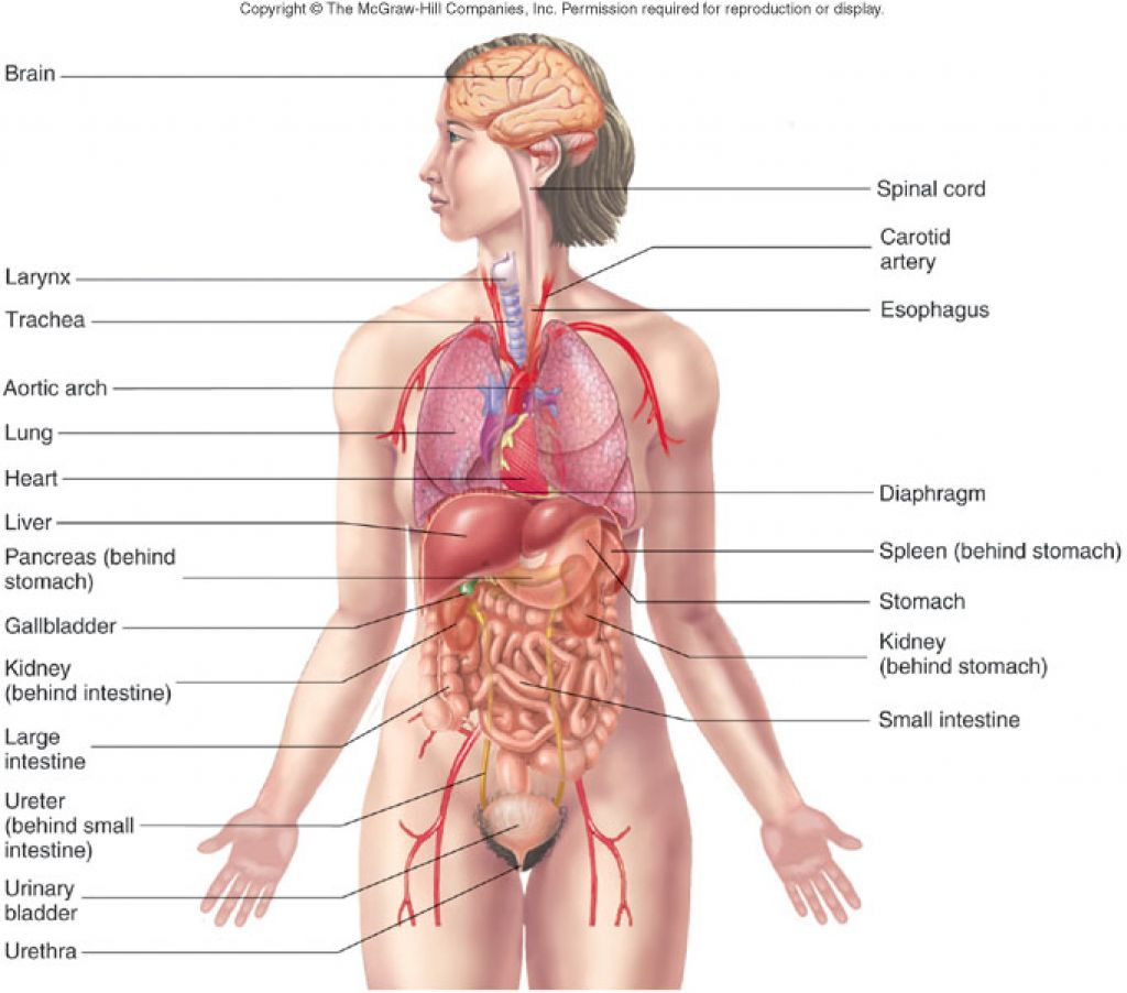
Free Human Body Organs, Download Free Human Body Organs png images, Free ClipArts on Clipart Library
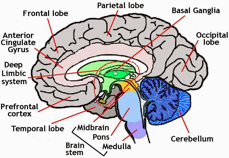
The Brain - Diagram and Explanation A diagram of how the brain works From Building Mental Muscle Glossary of Terms Six Brain Functions AMYGDALA: Lying deep in the center of the limbic emotional brain, this powerful structure, the size and shape of an almond, is constantly alert to the needs of basic survival including sex, emotional reactions such as anger and fear.
Illustrations of the Brain | Felix Song, M.D. // Interventional Neuroradiologist
Major Structures and Functions of the Brain - NCBI Bookshelf Visual functions occupy the occipital lobe, the bulge at the back end of the brain. The primary area for visual perception is almost surrounded by the much larger visual association area. Nearby, extending into the lower part of the temporal lobe, is the association area for visual memory —a specialized area in the cortex.

Parts of the Human Brain | Anatomy & Function - Study.com The parts of the brain include the cerebrum, the cerebellum, the brain stem, and the pituitary gland. The brain structure is protected by the skull, which is composed of the cranium and the bones...
Unlabeled Brain Diagram - Cliparts.co
Brain Anatomy and How the Brain Works - Hopkins Medicine The cerebellum ("little brain") is a fist-sized portion of the brain located at the back of the head, below the temporal and occipital lobes and above the brainstem. Like the cerebral cortex, it has two hemispheres. The outer portion contains neurons, and the inner area communicates with the cerebral cortex.

Anatomy and Functional Areas of the Brain | Doctor Stock
Brain Anatomy Labeled Stock Illustrations - Dreamstime How the eye works medical scheme poster, elegant and minimal vector illustration, eye - brain labeled structure diagram. Stylized and artistic medical design. Inner organ icons vector illustration collection set. Labeled medical and anatomical human brain, lungs, heart, liver and stomach. ... Labeled diagram with location and functions. Frontal ...

Detailed Labeled Diagram Of The Brain - Aflam-Neeeak
Anatomy of the Brain: Structures and Their Function Anatomy of the Brain. The anatomy of the brain is complex due its intricate structure and function. This amazing organ acts as a control center by receiving, interpreting, and directing sensory information throughout the body. The brain and spinal cord are the two main structures of the central nervous system. There are three major divisions of ...

Educative diagrams: The Female Reproductive System
Diagram Of Brain with their Labelings and Detailed Explanation A well-labelled diagram of a human brain is given below for further reference. Structure And Function Of The Human Brain Parts Of The Human Brain The human brain is divided into three main parts: Forebrain. Midbrain. Hindbrain. These three main parts comprises many small parts. Forebrain The forebrain is also called as Prosencephalon.
Brain Diagram Labeled Simple ~ DIAGRAM
Labeled Diagrams of the Human Brain You'll Want to Copy Now Labeled Diagrams of the Human Brain Central Core The central core consists of the thalamus, pons, cerebellum, reticular formation and medulla. These five regions are the central areas that regulate breathing, pulse, arousal, balance, sleep and early stages of processing sensory information.
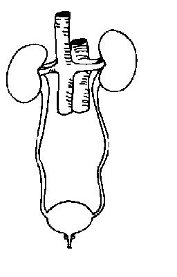
The Anatomy and Physiology of Animals/Excretory System Worksheet - WikiEducator
Labeled Brain Model Diagram - Science Trends The cerebrum is the largest and most complex portion of the human brain. The cerebrum's function is to control our actions and thoughts, either conscious or unconscious, and responses to stimuli. The cerebrum itself is typically divided into four different lobes: the temporal lobe, the parietal lobe, the occipital lobe, and the frontal lobe.
Post a Comment for "43 diagram of the brain with labels and functions"