40 brain pictures and labels
Human Brain Photos and Premium High Res Pictures - Getty Images 27,560 Human Brain Premium High Res Photos Browse 27,560 human brain stock photos and images available, or search for human brain anatomy or human brain illustration to find more great stock photos and pictures. of 100 NEXT Parts of the brain: Learn with diagrams and quizzes - Kenhub Labeled brain diagram First up, have a look at the labeled brain structures on the image below. Try to memorize the name and location of each structure, then proceed to test yourself with the blank brain diagram provided below. Labeled diagram showing the main parts of the brain Blank brain diagram (free download!)
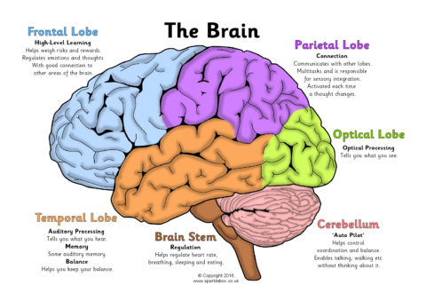
Labeled Brain Model Diagram - Science Trends The cerebrum is the largest and most complex portion of the human brain. The cerebrum's function is to control our actions and thoughts, either conscious or unconscious, and responses to stimuli. The cerebrum itself is typically divided into four different lobes: the temporal lobe, the parietal lobe, the occipital lobe, and the frontal lobe.

Brain pictures and labels
Brain Label (Remote) - The Biology Corner Brain Label (Remote) Shannan Muskopf December 29, 2020. This brain labeling activity was created for remote learners as an alternative to the labeling and coloring worksheet we would traditionally do in class. Instead of coloring and labeling on printouts, students use google slides to drag labels to the images or type the answers into text boxes. Human Brain Stock Photos, Pictures & Royalty-Free Images - iStock Browse 224,172 human brain stock photos and images available, or search for human brain anatomy or human brain illustration to find more great stock photos and pictures. Human brain icon in line style. Human brain icon in line style. For your design, logo. Vector illustration. Hot topics: Brain control of exercise, fatty fruit helps the ... May 22, 2022 · Hot topics: Brain control of exercise, fatty fruit helps the heart, sugary drinks and nutrition labels, artificial sweeteners and cancer risk, nostalgia for pain relief
Brain pictures and labels. 75,682 Brain Anatomy Stock Photos and Images - 123RF Brain Anatomy Stock Photos And Images 75,682 matches Page of 757 Brain lobes vector illustration. Human brain infographic vector. Brain lobes functions Serotonin pathway. Humans brain with serotonin pathways. psychiatric and neurological disorders. 3D render of a medical image showing male figure with brain tumour Neocortex vector illustration. Human Brain Diagram Photos and Premium High Res Pictures - Getty Images 1,000 Human Brain Diagram Premium High Res Photos Browse 1,000 human brain diagram stock photos and images available, or start a new search to explore more stock photos and images. of 17 NEXT Human Brain Diagrams and Detailed Information - Innerbody The brain needs to store many different types of information that it receives from the senses and that it develops through thinking in the association areas. Information in the brain is stored in a few different ways depending on its source and how long it is needed. Our brain maintains short-term memory to keep track of the tasks in which the ... Brain & Neuron Coloring Pages | Brain neurons, Neurons ... - Pinterest One shows internal brain structures, another shows the lobes of the brain and the third shows the important areas of the cortex. This large format provides: 1) enough room to record descriptions of all the functions of the parts 2) three clear diagrams to color 3) three fun foldables to use as all-in-one study guides for the brain.
Drawing Of The Brain With Labels - Painting Valley We collected 36+ Drawing Of The Brain With Labels paintings in our online museum of paintings - PaintingValley.com. ADVERTISEMENT LIMITED OFFER: Get 10 free Shutterstock images - PICK10FREE brain human diagram labeled anatomy label system easy physiology infant coronal neat nervous spinal simple cord rat Brain Diagram Labele... 633x512 41 0 Neuriva Plus Brain Performance Oral: Uses, Side Effects ... Neuriva Plus Brain Performance 1.7 Mg-400 Mcg-2.4 Mcg Capsule Not Applicable - Uses, Side Effects, and More Generic Name(S): B6-folic-B12-coffee-phosphatid View Free Coupon Illustration Picture of Brain Anatomy - Brain - eMedicineHealth Medical Illustrations Picture of Brain The brain is the complex organ responsible for processing sensory information (sound, touch, taste, sight, and smell). The brain controls voluntary and involuntary movements. Signals from the brain tell muscles to contract. Input from the brain controls the function of other organs in the body. Brain diagram with labels Images, Stock Photos & Vectors Find Brain diagram with labels stock images in HD and millions of other royalty-free stock photos, illustrations and vectors in the Shutterstock collection.
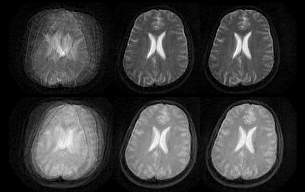
1,000+ of the Best Brain Pictures for Free [HD] - Pixabay 1,000+ of the Best Brain Pictures for Free [HD] 1,000 Pictures of Brain in HD Related Images: people human nervous system mind Pick the perfect brain picture for your project. HD to 4K quality, available for free on all devices! Brain MRI: How to read MRI brain scan | Kenhub Reading time: 20 minutes. Normal brain MRI. A brain MRI is one of the most commonly performed techniques of medical imaging. It enables clinicians to focus on various parts of the brain and examine their anatomy and pathology, using different MRI sequences, such as T1w, T2w, or FLAIR. MRI is used to analyze the anatomy of the brain and to ... Frontiers | 101 Labeled Brain Images and a Consistent Human Cortical ... We selected 101 T 1-weighted brain MR images that are: (1) publicly accessible with a non-restrictive license, (2) from healthy participants, (3) of high quality to ensure good surface reconstruction, and (4) part of a multi-modal acquisition ( T 2*-weighted, diffusion-weighted scans, etc.). Brain: Atlas of human anatomy with MRI - e-Anatomy - IMAIOS Anatomy of the brain (MRI) - cross-sectional atlas of human anatomy. The module on the anatomy of the brain based on MRI with axial slices was redesigned, having received multiple requests from users for coronal and sagittal slices. The elaboration of this new module, its labeling of more than 524 structures on 379 MRI images in three different ...
doctor sabelotodo: IVAN SHISHKIN
Brain labels Images, Stock Photos & Vectors | Shutterstock Find Brain labels stock images in HD and millions of other royalty-free stock photos, illustrations and vectors in the Shutterstock collection.

The Ever-Changing Brain – Daniel G Wood
Nervous System - Label the Brain - TheInspiredInstructor.com This brain part controls thinking. This brain part controls balance, movement, and coordination. This brain part controls involuntary actions such as breathing, heartbeats, and digestion. This part of the nervous system moves messages between the brain and the body. This part of the cerebrum interprets and sorts information from the senses.

WMU Psychology Department: Lisa Baker
Split-Brain: What We Know Now and Why This is Important for ... May 12, 2020 · For instance, Pinto et al., 2017a) found that the split-brain patient was much better at matching pictures to sample stimuli in the left visual field. Yet, for the exact same stimuli matching pictures to verbal labels was vastly superior when the stimuli appeared in the right visual field. Crucially, response type did not play any role.
MRIIT - Home
Diagram Of Brain with their Labelings and Detailed Explanation A well-labelled diagram of a human brain is given below for further reference. Structure And Function Of The Human Brain Parts Of The Human Brain The human brain is divided into three main parts: Forebrain. Midbrain. Hindbrain. These three main parts comprises many small parts. Forebrain The forebrain is also called as Prosencephalon.
Pineal Region Approach | Neuroanatomy | The Neurosurgical Atlas, by Aaron Cohen-Gadol, M.D.
Functional network organization of the human brain - PMC Nov 17, 2011 · Auditory-face correlations are significantly higher than auditory-hand correlations in both cohorts (p < 0.001, two-sample two-tail t-test). Bottom, slices from the 4% tie density modified voxelwise analysis, with labels on relevant thalamic nuclei (numbers are z coordinates).
Posterolateral View of the Suboccipital Region | Neuroanatomy | The Neurosurgical Atlas, by ...
Brain labelled Images, Stock Photos & Vectors | Shutterstock Find Brain labelled stock images in HD and millions of other royalty-free stock photos, illustrations and vectors in the Shutterstock collection.

brain labeled
Brain: Anatomy, Pictures, Functions, and Conditions The Brain Stem. PIXOLOGICSTUDIO/SCIENCE PHOTO LIBRARY / Getty Images. The brainstem is an area located at the base of the brain that contains structures vital for involuntary functions such as the heartbeat and breathing. The brain stem is comprised of the midbrain, pons, and medulla. 3.

Sain Creationz: STOP Torture!
687 results for labeled brain in images - Adobe Stock Search from thousands of royalty-free Labeled Brain stock images and video ... Central Organ of Human Nervous System Brain Frontal Lobe with Labels Anatomy.

Label Brain Quiz
Labeled Diagrams of the Human Brain You'll Want to Copy Now Labeled Diagrams of the Human Brain Central Core The central core consists of the thalamus, pons, cerebellum, reticular formation and medulla. These five regions are the central areas that regulate breathing, pulse, arousal, balance, sleep and early stages of processing sensory information.

Wheels Are Everything: 1966 Chevrolet C10
Human Brain Anatomy - Components of Human Brain with Images ii. Cerebellum—the Sub-Cerebral Region: Placed under the cerebrum, it is a relatively smaller component of brain. Cerebellum is assigned the task of controlling and coordinating the movement of muscles, and the maintenance of balance and body posture. iii. Brainstem: Medulla + Pons + Midbrain. YouTube.
Label the Brain Worksheets
Human Brain Worksheets - Superstar Worksheets Human Brain Worksheets valerie 2021-12-20T10:23:35-08:00. Learning about the human brain is a fascinating area of study for students of all ages. Use these free printable human brain worksheets for your science notebooks, journals, and science projects.

Brain Labeling Game - Made By Creative Label
Brain label Images, Stock Photos & Vectors | Shutterstock Find Brain label stock images in HD and millions of other royalty-free stock photos, illustrations and vectors in the Shutterstock collection.

Blank Brain Diagram To Label - Hanenhuusholli
101 Labeled Brain Images and a Consistent Human Cortical Labeling ... We selected 101 T 1-weighted brain MR images that are: (1) publicly accessible with a non-restrictive license, (2) from healthy participants, (3) of high quality to ensure good surface reconstruction, and (4) part of a multi-modal acquisition ( T 2*-weighted, diffusion-weighted scans, etc.).

Which Gets More People To Quit Smoking, Graphic Images On Cigarette Packs Or The Surgeon General ...
Human brain with labels, illustration Stock Photo - Alamy Download this stock image: Human brain with labels, illustration. - PHBYR3 from Alamy's library of ... Pre-pay for multiple images and download on demand.

Erin Faye Jasiura _ illustration portfolio
Haloperidol Oral: Uses, Side Effects, Interactions, Pictures ... Haloperidol is also used for severe behavior problems in hyperactive children when other treatments or medications have not worked.Haloperidol is a psychiatric medication (antipsychotic-type) that ...

33 Human Brain With Label - Labels Database 2020
Anatomy of the Brain | Simply Psychology The occipital lobes are located at the back of the brain behind the temporal and parietal lobes and below the occipital bone of the skull (Figure 7).. The occipital lobes receive sensory information from the retinas of the eyes which is then encoded into different visual data. Some of the functions of the occipital lobes include being able to assess size, depth, and distance, determine colour ...
Post a Comment for "40 brain pictures and labels"